I’m grateful to those people and organisations mentioned below for making the amazing images used in the banners on this website freely available! (All other images are from stock photo websites).

Vascular flows in the human brain imaged using ultrasound localisation microscopy and Iconeus functional ultrasound equipment. Image © EPSCI Paris from a video published on YouTube, and relating to work described in the following research paper: C. Demené, J. Robin, A. Dizeux, B. Heiles, M. Pernot, M. Tanter and F. Perren, Transcranial ultrafast ultrasound localization microscopy of brain vasculature in patients, Nature Biomedical Engineering, 2021, 5: 219–228, http://doi.org/10.1038/s41551-021-00697-x.
Microtome stem section of Lettuce-leaf Blumea (Blumea lacera) from the family Asteraceae, generated by microtome slicing and using safranin staining. Original image available here. Credit: himmaliyah, CC BY-SA 4.0, via Wikimedia Commons.
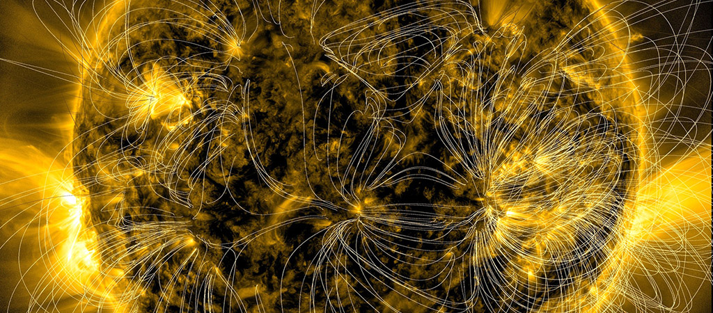
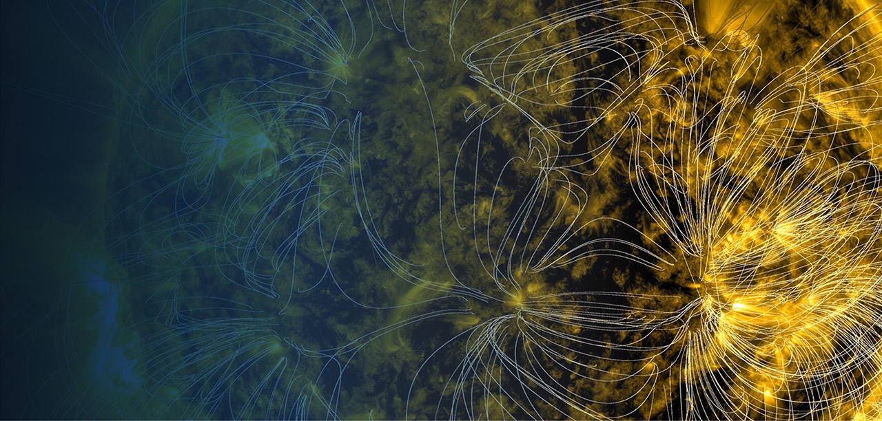
Magnetic field lines obtained over a period of four days, overlaid on an image of the sun. Original image available here. Credit: NASA Goddard Space Flight Center from Greenbelt, MD, USA, Public domain, via Wikimedia Commons.
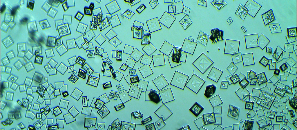
Microcrystals of cesium perchlorate (CsClO4) at 40× magnification, prepared by mixing cesium nitrate and sodium perchlorate. Original image available here. Credit: Arctur.rd, CC BY-SA 4.0, via Wikimedia Commons.
Close up of printed circuit board. Original image available here. Credit: Harland Quarrington/MOD, OGL v1.0OGL v1.0, via Wikimedia Commons.
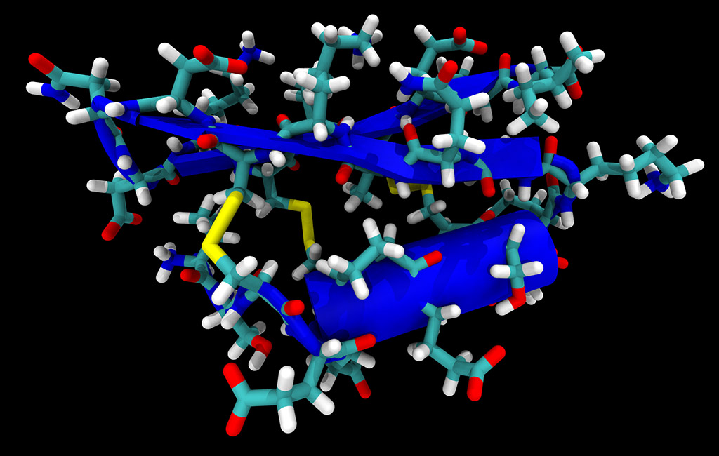
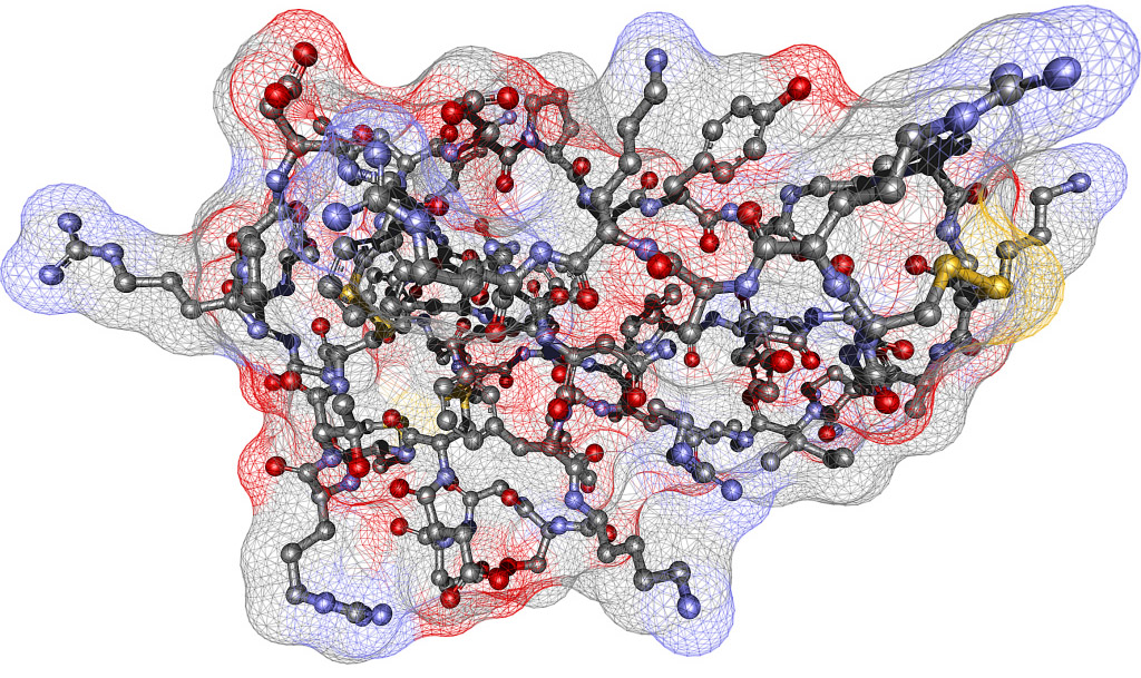
Structure of neurotoxin P01 from the Fat-tailed Scorpion (Androctonus mauretanicus mauretanicus), using data from the Protein Data Bank (ref. 1ACW). Original image available here, with cited reference: E. Blanc et al., Solution structure of P01, a natural scorpion peptide structurally analogous to scorpion toxins specific for apamin-sensitive potassium channel, Proteins, 1996, 24: 359–369, https://doi.org/10.1002/(SICI)1097-0134(199603)24:3<359::AID-PROT9>3.0.CO;2-B. Credit: Ayacop, Public domain, via Wikimedia Commons.
Kinetic simulation of the plasma in an accretion disk for a neutron star or black hole, showing the multi-scale structure of the magnetic turbulence. Original image available here. Image credit: Giannandrea Inchingolo, CC BY-SA 4.0, via Wikimedia Commons.
Polarised light microscope image of crystallisation of molten sulfur. Original image available here. Image credit: AlexmarPhoto, CC BY 4.0, via Wikimedia Commons.
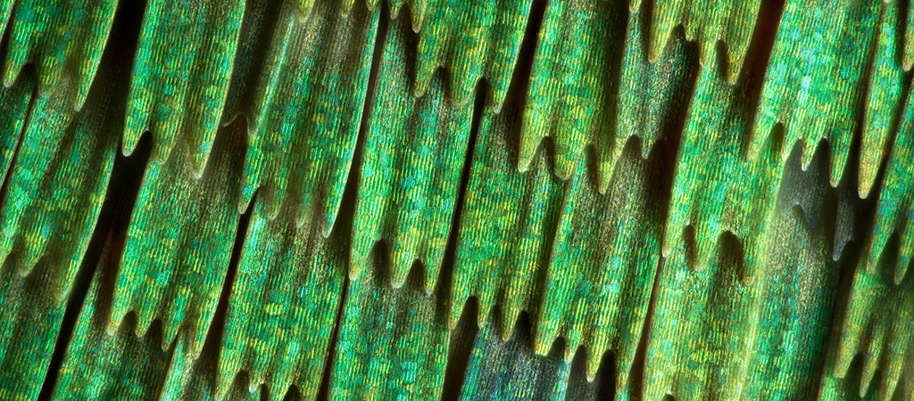
Wing scales of the Green Hairstreak butterfly (Callophrys rubi), at 50× magnification and using focus stacking. Original image available here. Image credit: Mikron86, CC BY 4.0, via Wikimedia Commons.
Multi-walled carbon nanotubes prepared by passing methane over a magnetite substrate at a temperature of 1100 °C. Original image available here. Image credit: Stanislav.nevyhosteny, CC BY 4.0, via Wikimedia Commons.
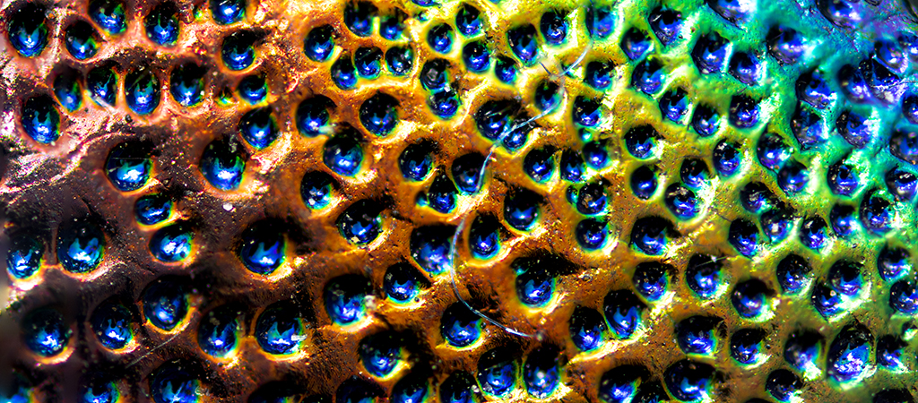
Structural colour in the cuticle of the beetle Cheloderus penai, caused by light interference within the alternating nanometre-scale layers of chitin. Original image available here. Image credit: Brandon Antonio Segura Torres & Priscilla Vieto Bonilla, CC BY-SA 4.0, via Wikimedia Commons.
Ball-and-stick model of bovine trypsin inhibitor, visualised without hydrogen atoms and using data from the Protein Data Bank (ref. 2PTC). Original image available here. Image credit: Anmoll at de.wikipedia, CC BY-SA 2.0 DE, via Wikimedia Commons.
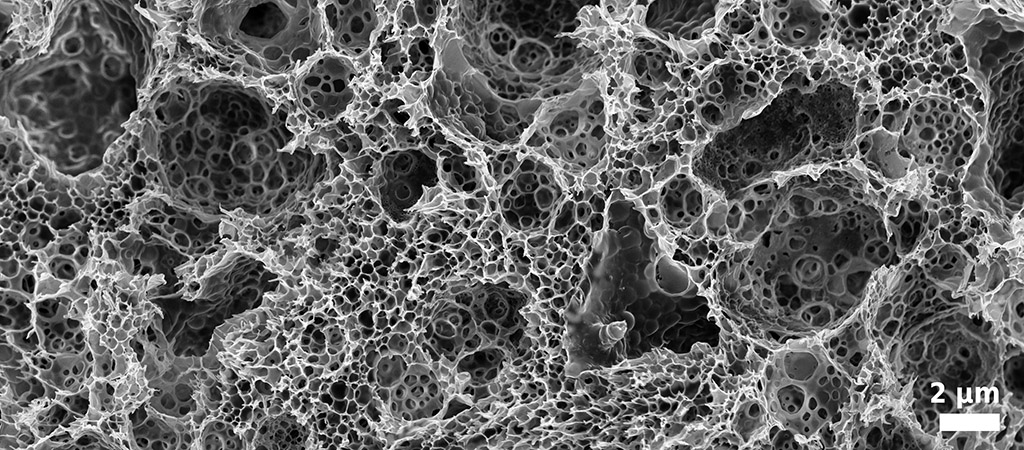
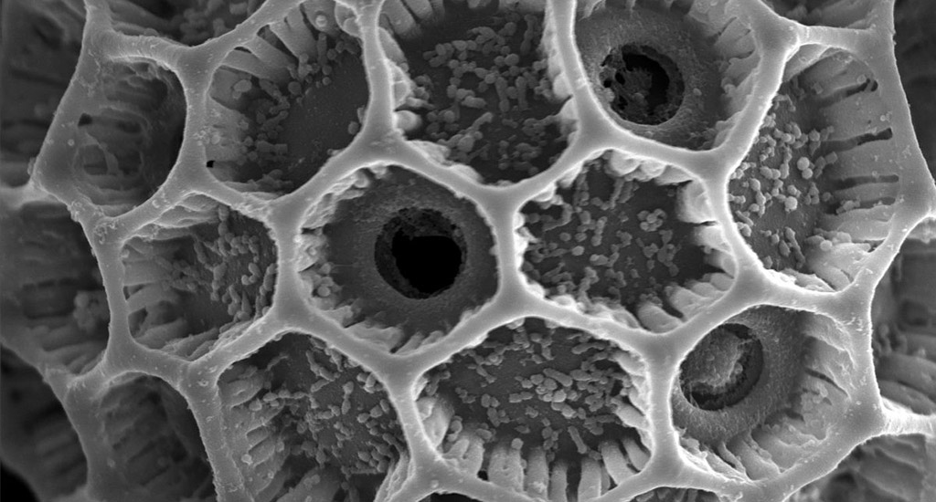
Scanning electron microscopy image of biodegradable polymer implant during hydrolysis, obtained by field-emission scanning electron microscopy. Original image available here. Image credit: Julia Higuchi, Jan Mizeracki, CC BY 4.0, via Wikimedia Commons.
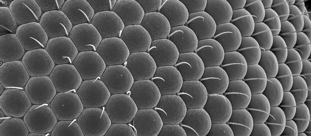
Scanning electron microscopy image of the compound eye of a fungus gnat (family Mycetophilidae). Original image available here. Image credit: Pavel kejzlar, CC BY 4.0, via Wikimedia Commons.
Scanning electron microscopy image of pollen of Cup-and-saucer Vine (Corbaea scandens). Original image available here. Image credit: Marie Majaura, CC BY-SA 3.0, via Wikimedia Commons.
Scanning electron microscopy image of a fractal pyramid with a base of length 141 µm, produced by two-photon polymerisation. Original image available here. Image credit: Spanholz, CC BY-SA 4.0, via Wikimedia Commons.